
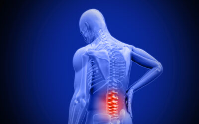
Spinal stenosis refers to abnormal narrowing of the hollow neural passageways running through the articulating vertebral bones of the spine. This constriction of space leads to compression, inflammation, and damage to the intricate nerves controlling sensation, muscle activity, organ function, and reflexes below the sites affected. Understanding key causal contributors helps guide appropriate diagnosis and management.
Spine Anatomy and Spinal Stenosis
The spinal cord connects to the brain, descending through bony tunnels formed by the stacked vertebrae comprising the flexible spine to relay bidirectional communication with body areas it supplies via branching nerves. Maintaining adequate patency of this conduit proves essential for normal mobility and health, as compression incites neurological dysfunction.
The neck and low back spinal regions undergo the highest mechanical stresses in day-to-day movement, making them most prone to developing consequential stenosis – but causative factors arise throughout all spinal levels over decades of use.
Major Causes
Various spinal problems ultimately narrow the delicate neural space available, producing inflammation and characteristic symptoms in zones connected to afflicted nerve structures.
Gradual Degenerative Factors
As we age, expected “wear-and-tear” on the spine builds up over years of use. Flexible tissues get exhausted from bearing weight and bending daily. Slowly, their stretchiness declines until they touch during normal movement, pressing on pain nerves. Spinal stenosis from aging happens bit-by-bit as disks, joints, and ligaments lose springiness that kept open spaces. No sudden injuries cause it. Years of everyday activity like sitting, walking and lifting take a toll on their cushioning. Older patients mostly develop narrowing canals due to predictable aging effects rather than specific events. Keeping flexible tissues resilient for as long as possible slows this gradual closure process.
Disc Breakdown
Intervertebral discs dry out over time, losing hydration elements that allow their gel-filled centers to withstand compression between vertebral bones. The discs thin and flatten, forcing vertebrae to rotate closer together. Declining thickness distributes vertical forces abnormally onto posterior joints linking each spinal segment instead of cushioning impact. This strains articulations not designed to bear as much weight.
Facet Joint Arthrosis
Increased mechanical loads wearing down fibrocartilage facet joint surfaces permits direct contact between inflammatory bone tissue. Fluid stretches joint capsules outward while bony heaps develop along meeting borders trying to stabilize their connections– which narrows exiting lateral spinal spaces. Inflamed synovium also secretes degradative enzymes thinning stabilizing spinal ligaments.
Flakey Buckled Ligaments
The ligamentum flavum lines the back portion of the spinal canal. This important ligament lacks normal blood flow. Constant pressure causes its tissue to start breaking down over time. Broken down material can trigger swelling while becoming less flexible and stretchy. Parts of the ligament may also turn into bone that then protrudes into the hollow space needed for nerves and fluid flow. The swollen, stiffened ligament can bulge into critical areas, contributing to symptoms.
Bone Spur Development
As the spine loses stability from damage and wear over time, the body tries to compensate by having bone overgrow along the edges of vertebrae and joints. This extra bone is meant to provide more support, but it can protrude into spaces needed for nerves. Bony spurs arising from the outer bone layers to reinforce the crumbling inner bone also creep into openings where nerves exit the spine. These bony protrusions stiffen flexibility in the spine. They may rub directly against or compress nerve fibers, causing inflammation and pain.
Congenital Factors
Some people are born with spine problems that raise their risk for stenosis symptoms down the road – even with a healthy lifestyle. They might have naturally narrow canal space or unusual bone shapes. Having less room to start means added growth or injury more easily presses on nerves. So kids diagnosed with conditions like Morquio syndrome or achondroplasia often face teenage or adult spinal problems. Their minimal extra capacity leaves little padding before nerve contact as the spine ages. Staying ahead of changes means hopefully preventing major issues. But these patients need extra monitoring since they lack wiggle room for developing narrowing before feeling effects. Keeping a close eye out allows treating emerging symptoms early.
Previous Injury
Injury causes sudden stenosis flares rather than slowly worsening over many years. Events like falls, car accidents, sports hits, or blasts over-compress spine bones and tissues. Exceeding safe loads – like from heavy overhead lifting – can also damage the spine’s ability to handle force.
When delicate nerves get crushed beyond their flexibility, bleeding and swelling rapidly fill in space meant just for fluid and nerves. Even surgery to stabilize injured segments leaves scar tissue or hardware that crowds nerve openings. Spines that endure trauma face higher stenosis risk long-term, even after bones heal.
Sudden compressing traumas initiate inflammation occupying previous open areas suited only for fluid and nerves, unlike slow arthritis closure. So recovering patients require extra monitoring to treat nerve compression signs early before severe issues develop. Staying ahead of post-traumatic stenosis gives the best outcomes.
Spinal Tumors
Abnormal tissue growths arise within the cord itself or inside adjacent vertebral bones in some cases. Tumors may be metastatic spread from cancers like breast, lung, prostate or benign pockets of defective cell clones forming neoplasia like neurofibromas or meningiomas along delicate membranes. Since even smaller masses substantially fill the central hollow canal, symptoms manifest early. Nerves suffer damage when unable to transpose around enlarging lesions.
Spinal Infection / Abscess
Infections inside the spine also create swelling that crowds nerves. Bacteria disc infections or shingles viruses attack nerve roots and tissue layers around the spinal cord. Pus buildup distorts anatomy. Floods of immune cell waste, swelling chemicals, and excess fluid drown once open areas. Bad infections easily spread to distant sites after entering the spine’s immune-protected channels within bones. Unlike slow arthritis closure, sudden spine infections severely damage nerves. Quickly treating contamination and drainage becomes crucial before permanent paralysis or disability develops. Restoring stability after treating the infection requires carefully rebuilding anatomy planes and monitoring nerve recovery.
Non-Spine Generators
Less commonly, non-spine origins falsely blamed for signs of stenosis instead refer pain into spine areas from remote sites. Common offenders include piriformis syndrome, sacroiliac joint dysfunction, hip osteoarthritis, or vascular perfusion diseases like peripheral artery stenosis. To know if the spine bones themselves are causing compression, other possible causes first need to be ruled out.
Symptoms Arising from Stenosis
As inflammation and narrowing happen in the hollow spaces down the center of the spine, it can affect both nerve signaling and fluid flow. Nerves exiting from areas of narrowing will then cause symptoms in the body parts they connect to. Patients describe matched pain, numbness, tingling, weakness, cramping, heavy sensations, or balance issues. Exact symptoms depend on spinal level and amount of nerve compression.
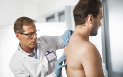
Diagnostic Steps
There are a multitude of conditions that can cause similar spine or nerve problems. Doctors need specific tests and scans to correctly pinpoint if spinal stenosis is behind the nerve compression. They start by asking about symptoms and doing hands-on physical checks for clues matching stenosis. Then MRI’s are ordered to closely map soft tissues along the spine segments in question. MRIs show nerve issues plus swelling and redness taking up space. By linking patients’ nerve function losses and inflammation areas to abnormal scan findings, doctors make an accurate stenosis diagnosis. They can zone in on what structures make canals narrow. Matching up descriptions, exam findings and imaging results is how doctors confirm spinal stenosis causes compression versus other possible conditions.
Treatment Options
Once testing shows what is narrowing the spinal canals, treatment focuses on easing the compression and swelling. Mild cases might get better with rest, medication, shots, braces, or physical therapy. However, many people need surgery to widen bony canals if pain and weakness do not improve after exhausting other options.
The main goal is protecting nerve function with spinal stenosis. Figuring out what structures are involved allows choosing treatments to provide real relief that sticks. While narrowing cannot be undone, slowing the progression and keeping symptoms in check helps patients stay active and feel their best. Staying on top of options to reduce compression and calm inflammation means those living with this condition can keep enjoying life on their own terms.
Conclusion
In summary, spinal stenosis refers to gradual narrowing of the hollow nerve network running through the spine bones. Delicate nerves pass through these canals. As space tightens, nerves get compressed, causing symptoms. Understanding what spine changes cause swelling and pinching allows matching scans to patient signs. After an accurate stenosis diagnosis, treatment focuses on easing the nerve compression and controlling inflammation. Options include medications, therapy, braces, shots or surgery to widen bony canals if other efforts do not protect nerve function. The goal is calming irritation around narrowed spaces and preserving nerve health to manage symptoms. Keeping an eye on how spinal stenosis progresses and finding the right treatments for nerve issues are important.

Written by Dr. Tony Mork
Orthopedic Spine Surgeon
I’m Dr. Tony Mork, MD, a Minimally Invasive Orthopedic Spine Surgery Specialist in Newport Beach, California. With over 40 years of experience, I’m dedicated to providing information for all topics that involve neck and back pain.
February 5, 2024
Contact a Spine Specialist Near You!
Related Articles
Lower Back Pain | An Overview
Lower back pain is an exceedingly common affliction that affects up to 80% of adults at some point in their lives. Understanding the underlying causes of lower back pain is key to finding the appropriate treatment and relief. Anatomy of the Lower Back The lower back,...

Degenerative Disc Disease | Causes and Risk Factors
Degenerative disc disease describes age-related wear and tear to the rubbery spinal discs cushioning the vertebrae that can lead to chronic back or neck pain. While partly genetic, there are also lifestyle factors and injuries that accelerate disc degeneration over...
Upper (Thoracic) Back Pain | What is Causing My Pain?
Upper back pain is a surprisingly common affliction, affecting up to one-quarter of the adult population at some point. While not as prevalent as lower back pain, discomfort in the thoracic region still accounts for a substantial slice of musculoskeletal complaints...