
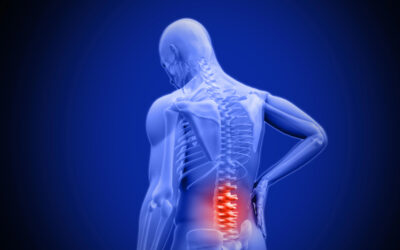
Spinal stenosis refers to abnormal narrowing of the spinal canal that causes compression of the spinal cord or spinal nerves traveling through the vertebral column to the rest of the body. The spinal cord extends down from the brain, carrying nerve signals and motor control pathways to and from the trunk and limbs. As the narrowing leads to inflammation or impingement of this delicate tissue, patients experience neurological symptoms and pain in the neck or back along with connected areas.
Anatomy of the Spine
Understanding spinal stenosis requires some knowledge of basic spine anatomy. The spine forms the flexible central support structure that allows the body to stand upright and move in various directions. This column consists of 33 total vertebrae, separated into 5 regions – cervical, thoracic, lumbar, sacral, and coccygeal.
The cervical section contains the first 7 vertebrae beginning immediately under the skull. Twelve thoracic vertebrae make up the upper and mid back area. Next in sequence are 5 larger, weight-bearing lumbar vertebrae in the lower back. The sacrum consists of 5 fused vertebrae fixed between the hip bones, and finally 4 tiny fused vertebrae form the tailbone or coccyx.
Vertebrae
The spine is made up of individual bones called vertebrae. Each vertebra has a front segment that frames the hollow spinal canal space where nerves pass through. It also has a back portion called the neural arch. The neural arches link by facet joints to the vertebrae above and below, forming the continuous spine. The arches have projections of bone that serve as attachment points for muscles. Together, these arched structures protect the spinal cord.
Between the vertebral bodies lie intervertebral discs. These act as cushions, allowing slight movement, bending and weight distribution along the spine. Their flexibility is important for mobility in the human body. However, they can be susceptible to damage over time.
Spinal Canal and Nerves
On the back side of the spine, the stacked vertebral arches contain a hollow column housing the spinal cord bundle of nerves running from neck down to low back. At each vertebra, nerve roots branch out right and left through small openings, connecting the spinal cord signals to large peripheral nerves that relay messages throughout the body.
It’s crucial to maintain enough room for the spinal cord and those transverse nerve branches despite frequent bending motions and injury over a lifetime. This space allows normal signal relay between brain and body which makes mobility and function possible. Loss of space puts critical nerves at risk.
Types of Spinal Stenosis
Abnormal narrowing of the nerve pathways can happen in several areas within the complex spinal anatomy. Identifying the exact location of compression helps determine what’s causing it and guide the right treatment methods. The main categorized types are central stenosis, foraminal stenosis, and lateral recess stenosis.
Central Spinal Stenosis
This common degenerative type involves narrowing of the overall central canal containing the spinal cord and cerebrospinal fluid. Enlarged facet joints, bulging discs, bone spurs, thickened ligaments, or spinal tumors infringe into the space available for these central soft tissue structures. The cord itself becomes compressed or inflamed, causing neurological problems in the areas it supplies below the compression site along the spine.
Foraminal Stenosis
The nerve roots need to pass through small openings between vertebrae called neural foramina. This allows them to exit the spinal canal and reach the arms, legs, and other areas. Foraminal stenosis means these openings narrow, specifically where the nerves are trying to exit. Bone spurs or arthritic overgrowth of the facet joints often cause the compression. When inflammation and swelling occurs in the already narrowed spaces, it pinches the nerves trying to pass through. Foraminal stenosis most often affects just one side at a time.
Lateral Recess Stenosis
The lateral recesses are small tunnels on each side of the spinal canal. This is where nerves first exit the cord before entering the openings between vertebrae. Like other stenosis types, anything pressing on this nerve path can cause symptoms. Most often, enlarged facet joints create issues in the lateral recesses.
Stenosis can happen in the neck, middle, or lower spine. But lumbar stenosis leading to leg pain and difficulty walking distances is especially common. Figuring out exactly where the narrowing and nerve compression occurs guides the precise diagnosis and best treatment options.
Symptoms of Spinal Stenosis
Patients with spinal stenosis experience neurological symptoms corresponding to inflammation or compression in areas connected to the central spinal cord itself or the branching peripheral nerves. Numbness, pain, tingling, weakness, and balance difficulties typically intensify when standing upright and ease somewhat while sitting or bending forward.
Lumbar Spinal Stenosis
In the lower back, symptoms often first show up in the legs, since irritated nerve roots exit here to supply the foot and calf areas. You may have leg cramping, heaviness, or pain when walking that goes away when you sit or lean forward – called neurogenic claudication. At least one foot likely feels tingling, numb, or painful eventually if untreated. Balance issues walking can also happen. In severe cases, bladder or bowel control problems could even occur if the spinal cord gets compressed.
Cervical Spinal Stenosis
When narrowing and nerve compression happens in the neck vertebrae, symptoms usually radiate down the arms rather than the legs. You may experience numb, painful hands with clumsiness and loss of fine finger control. This can interfere with normal tasks like writing or typing. Neck discomfort when holding the head upright commonly builds as nearby nerve irritation inflames muscles around the shoulders and upper back. Balance problems walking or repeated tripping can mark more advanced cases.
Causes of Spinal Stenosis
Later in life, gradual “wear and tear” on the spine like bone spur formation, disc problems, enlarged ligaments and facet joints prove the most common reasons for spinal canal narrowing. Degenerative changes slowly accumulate, stiffening spine flexibility and mobility. The small built-in buffer space gets occupied by inflammatory tissue or bone overgrowth, until symptoms emerge signaling nerve compression.
Other causes include congenital malformations making stenosis inevitable, traumatic fractures/dislocations abruptly compressing tissue beyond capacity, and spinal tumors or cysts bulging into the canal space. Understanding all contributing factors aids diagnosis.
Diagnosing Spinal Stenosis
Since other neurological conditions like peripheral neuropathy, sciatica from piriformis syndrome, vascular claudication in the legs, and multiple sclerosis also frequently enter the differential diagnosis when patients present with complaints of limb tingling, numbness, or balance problems, doctors utilize various imaging and laboratory testing to pinpoint spinal stenosis as the actual culprit. They combine insights from precise descriptions of symptom quality, timing, and aggravating factors with visualizing what structures explain internal compression along the vertebral levels in question.
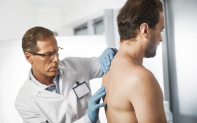
Physical Exam
Doctors specially trained in spinal conditions use particular examination techniques to help determine the cause of symptoms. They will observe how you move about the room, get up from a chair, balance, and walk. Your spine flexibility is checked by having you bend forward, extend back, and twist from side to side.
Signs like a wide stance, shakiness, limited spinal motion, tilted posture, or neck position changes can reflect bone contributions to nerve space crowding. Muscle strength testing identifies weakness suggesting impinged nerves. Reflex reactions help localize which nerve levels are affected. Sensation differences along nerve paths help find possible pinched nerves. Straight leg raising often recreates posterior thigh, leg or foot pain from compressed nerve roots in the low back.
Overall, the pattern of symptoms and exam findings seen with spinal stenosis differs enough from other spinal conditions to potentially allow an accurate diagnosis just based on the clinical assessment in many cases before ordering extra tests.
Imaging Studies
When doctors need anatomical views to directly link exam findings with visible nerve compression, imaging helps confirm diagnoses. X-rays detect fractured or misaligned bones but poorly show soft tissues. Myelograms use injected dye to coat the spinal cord and see its shape, but the needles risk more inflammation. MRIs safely distinguish tissues based on their water content without needles or radiation. Strong magnets create 3D reconstructions spotlighting disc herniations, swollen ligaments, bone spurs, tumors, and other canal crowding. CT scans finely portray abnormal bone and calcified deposits.
Comparing suspected affected areas against normal segments pinpoints pathological contributors explaining your symptoms. Additional electrical testing can check nerve conduction. Once imaging confirms areas of spinal narrowing, your doctor can connect these to clinical findings and guide appropriate treatments.
Treatments for Spinal Stenosis
When spinal narrowing causes mild symptoms, conservative nonsurgical options aim to manage inflammation and pain levels through medications, steroid injections, physical therapy, braces, and alternative treatments.
More advanced cases often require surgical intervention to provide lasting relief by removing tissue pressing on nerves. Common decompression procedures include laminectomy to cut away intruding bone and ligament and spinal fusion to stabilize the spine. Recently introduced spacers and devices can indirectly expand canal space without removing vertebral tissue.
Choosing the optimal treatments requires balancing severity of symptoms, degree of spinal instability, and expectations for improvement. Starting conservatively makes sense for most mild to moderate spinal stenosis given the risks and recovery accompanying invasive options. Rapidly worsening muscle weakness or bowel/bladder dysfunction indicates the need for expedited surgery to stop further nerve damage.
Living with Spinal Stenosis
Though spinal changes leading to stenosis are common with aging, many steps can ease the process and limit impacts. Prioritizing spine health through proper mechanics, suited exercise, and adaptive mobility helps retain function longer before surgery is needed. Identifying personal trigger activities reduces pain flares. Staying positive while proactively managing evolving symptoms lets many patients thrive for years after diagnosis by adapting rather than halting active lifestyles. There are options to smooth the transition towards stabilized, non-progressive stenosis.

Written by Dr. Tony Mork
Orthopedic Spine Surgeon
I’m Dr. Tony Mork, MD, a Minimally Invasive Orthopedic Spine Surgery Specialist in Newport Beach, California. With over 40 years of experience, I’m dedicated to providing information for all topics that involve neck and back pain.
February 5, 2024
Contact a Spine Specialist Near You!
Related Articles
Lower Back Pain | An Overview
Lower back pain is an exceedingly common affliction that affects up to 80% of adults at some point in their lives. Understanding the underlying causes of lower back pain is key to finding the appropriate treatment and relief. Anatomy of the Lower Back The lower back,...

Degenerative Disc Disease | Causes and Risk Factors
Degenerative disc disease describes age-related wear and tear to the rubbery spinal discs cushioning the vertebrae that can lead to chronic back or neck pain. While partly genetic, there are also lifestyle factors and injuries that accelerate disc degeneration over...
Upper (Thoracic) Back Pain | What is Causing My Pain?
Upper back pain is a surprisingly common affliction, affecting up to one-quarter of the adult population at some point. While not as prevalent as lower back pain, discomfort in the thoracic region still accounts for a substantial slice of musculoskeletal complaints...